Brain scans reveal two distinct biological profiles of migraine
A new analysis of brain imaging data suggests that migraines can be categorized into two biological groups based on brain structure and network connectivity, offering a potential new framework for understanding the condition.
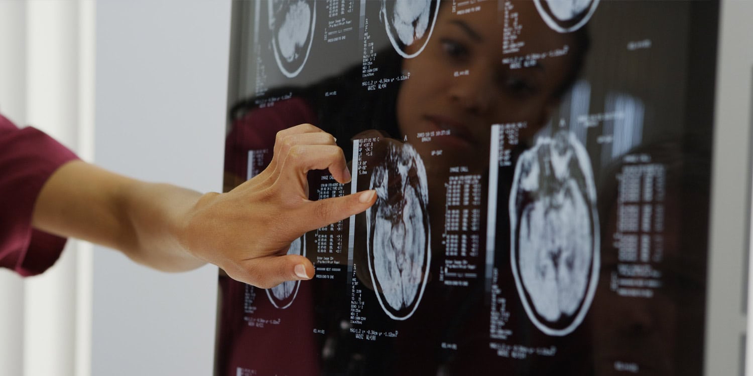
A recent study using brain scans has uncovered two separate biological profiles of migraine sufferers. These profiles are based on the unique wiring and structure of the brain, rather than the traditional symptom-based classifications. The research, published in Cephalalgia, suggests this new understanding could lead to more effective migraine treatments.
Migraine is a neurological disorder characterized by severe head pain, sensitivity to light, and other debilitating symptoms. Currently, doctors classify migraine based on the frequency of attacks and the presence of an aura – visual or sensory disturbances preceding the headache. However, this symptom-focused approach often fails to predict which treatments will be most effective for individual patients.
The researchers, led by Stanford University's Jaiashre Sridhar and Danielle D. DeSouza, aimed to identify biological subgroups using brain imaging techniques. They employed two types of MRI: structural MRI, which measures the physical dimensions of the brain, and functional MRI, which tracks blood flow to observe how different brain regions communicate when a person is at rest.
By analyzing brain scan data from 111 migraine sufferers and 51 healthy controls, the researchers identified two distinct biological subgroups. One group exhibited a higher burden of migraine, including longer duration headaches, reduced confidence in managing pain, and elevated connectivity between deeper brain structures and cortical networks responsible for attention, movement, and visual processing. This group also showed reduced brain volume in several cortical regions.
The second subgroup had a milder profile, with preserved brain structure and normal functional connectivity compared to the first group. When the researchers analyzed the data using only functional connectivity, the two subgroups remained consistent. However, when analyzing only structural MRI data, the initial groups diverged into entirely different clusters, indicating that functional connectivity drives most of the initial subgroupings.
Further sensitivity analysis using a finer-grained brain map produced consistent results, suggesting these biological groups are robust. However, the study's limitations include its single-timepoint data, inability to track migraine progression, and lack of information on preventive medication use. Additionally, the study did not align the biological findings with traditional migraine categories like chronic or episodic migraine.
Written by urgent.news from PsyPost's reporting — not their text. Machine-written — may contain errors; check the original before relying on it.
This story
This is one outlet's version. Read the fullest account.