How imaging offers a window to treat wake-up strokes
One in five ischaemic strokes is a wake-up stroke. “This means a person wakes up from his/her sleep with symptoms of stroke. The symptoms include facial drooping, difficulty speaking, sudden vision problems and weakness on one side of the body
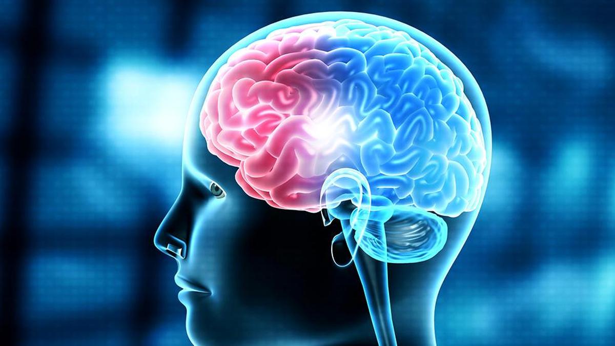
Strokes occurring during sleep, known as wake-up strokes, present unique challenges for medical professionals. Unlike regular strokes, the onset of symptoms in wake-up strokes is not always apparent until the patient wakes up, making it difficult to determine the precise timeline of the stroke's initiation. This lack of a clear onset time hinders doctors' ability to assess the extent of brain injury and the potential for salvageable tissue, which is crucial for administering prompt treatment.
Brain imaging plays a vital role in diagnosing and treating wake-up strokes. An immediate MRI brain scan or, if unavailable, a CT angiogram can provide critical information about the extent of brain damage and blood flow to various regions of the brain. These scans help identify diffusion-perfusion mismatch, indicating brain tissue that may still be salvageable through thrombolysis or mechanical thrombectomy.
The term "time is brain" still applies to wake-up strokes, emphasizing the urgency of swift diagnosis and treatment.
Neurologists like M. Radha, head of department and professor of neurology at Government Kilpauk Medical College (KMC) Hospital in Chennai, stress the importance of seeking immediate medical care upon noticing stroke symptoms, which may include facial drooping, difficulty speaking, sudden vision problems, and weakness on one side of the body. Within 4.5 hours of symptom onset, thrombolysis can be performed, but in wake-up strokes, the exact onset time remains unknown, making rapid action essential.
In cases of wake-up strokes, the lack of a defined onset time necessitates swift action based on imaging results. Dr. Radha highlights the significance of MRI imaging, particularly diffusion-weighted imaging and FLAIR (Fluid-Attenuated Inversion Recovery) imaging, as they can reveal salvageable brain tissue even without knowing the exact time of stroke onset.
Similarly, MRI perfusion or CT perfusion studies offer insights into salvageable brain tissue, guiding decisions on thrombolysis, mechanical thrombectomy, or conservative management.
The Tamil Nadu Accident and Emergency Care Initiative and Stroke Care and Rapid Intervention with Plasminogen activator and Thrombectomy (SCRIPT) program, implemented by the Tamil Nadu government, has streamlined treatment for wake-up stroke patients. Enhanced coordination between administration, emergency departments, radiology, and neurology departments has significantly reduced the door-to-needle time, ensuring timely intervention for patients with stroke symptoms.
Dr. Radha notes that increased awareness of stroke symptoms and improved referral systems contribute to better care outcomes.
Dr. Shubha Subramanian, senior consultant at Kauvery Hospitals, points out that individuals experiencing wake-up strokes may not initially recognize their symptoms, as they may have difficulty getting up or even fall upon waking. Family members play a crucial role in identifying these symptoms and seeking immediate medical assistance. The BE FAST acronym, which stands for Balance, Eyes, Face, Arm, Speech, and Time, serves as a useful tool to spot the warning signs of stroke.
Written by urgent.news from The Hindu - Sci-Tech's reporting — not their text. Machine-written — may contain errors; check the original before relying on it.
This story
This is one outlet's version. Read the fullest account.
- How imaging offers a window to treat wake-up strokes thehindu.com