Brain structure linked to how well patients respond to an antidepressant, small study finds
Why do some people with depression respond to medication while others don't? A small study points to the brain's outer layer, finding that the folding complexity and thickness of certain cortical regions tracked how much patients improved.
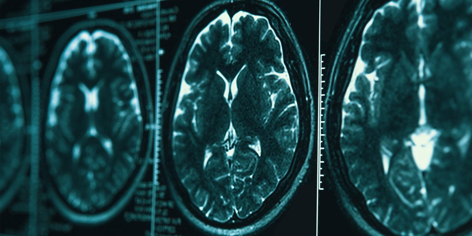
A new study reveals a potential link between the structure of certain brain regions and the effectiveness of an antidepressant called vortioxetine. Led by researchers at Beijing HuiLongGuan Hospital in China, the small study examined the brain scans of 22 people with major depressive disorder before and after they took the medication for about eight weeks. The researchers also compared the results with 21 healthy participants.
The study focused on the cortex, the brain's outer layer, measuring features such as cortical thickness, gyrification index (how much the cortex is folded), sulcus depth (depth of the grooves between folds), and fractal dimension (a measure of folding complexity). Participants with depression had less folding in the right insula compared to the healthy participants.
Greater fractal dimension, indicating more complex folding, in specific frontal and visual brain regions was associated with better symptom improvement. The frontal regions are involved in emotion regulation and executive control, while the visual region helped in recovery of feelings.
Shallower grooves in the left occipital cortex were linked to better memory improvement, specifically delayed memory recall. The researchers used a technique called leave-one-out cross-validation to check the consistency of these associations, taking into account participants' age and sex. The findings suggest that brain structure could potentially predict how well patients respond to vortioxetine treatment, offering a more personalized approach to care for depression.
Written by urgent.news from PsyPost's reporting — not their text. Machine-written — may contain errors; check the original before relying on it.