Miniature microprobe enables internal imaging of smaller blood vessels than ever before
A microprobe can navigate and image smaller arteries than previously possible, helping diagnose and plan treatments for vascular diseases The post Miniature microprobe enables internal imaging of smaller blood vessels than ever before appeared first on Physics World .
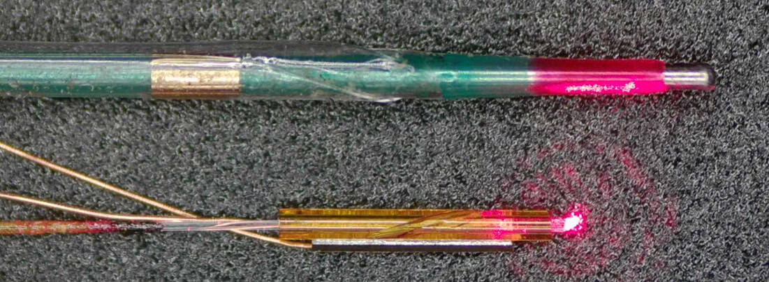
Cardiovascular disease is the leading cause of death worldwide, with atherosclerotic disease – resulting from fatty plaque build-up in arteries – accounting for approximately three-quarters of these fatalities. Intravascular optical coherence tomography (IV-OCT) is a modern imaging technique that enables detailed cross-sectional visualization of these fatty plaques, significantly improving atherosclerosis diagnosis and treatment planning.
A research team from China, led by Dawei Wu of Nanjing University of Aeronautics and Astronautics and Rui Liu from Nanjing University Medical School, has developed a miniature microprobe that pushes IV-OCT capabilities to smaller blood vessels and higher resolution. This innovative probe could potentially enable imaging of the cerebrovasculature – the network of tiny vessels within the brain.
The IV-OCT imaging technique relies on near-infrared light emitted and received at the end of a catheter to examine the walls and texture of blood vessels. By comparing the wavelength and phase offset of reflected light with reference light reflected from a mirror, and utilizing 360° sensor rotation, IV-OCT generates high-resolution images.
Traditionally, IV-OCT catheters measure around 2 mm in diameter and perform optimally in vessels of 10 mm in diameter, which limits their application to medium and large arteries. The newly developed probe, however, boasts a 0.55 mm diameter, making it ideal for imaging 2 mm vessels while maintaining full 360° field-of-view.
Key to the probe's small size and superior imaging capabilities is its unique piezoelectric micro-optical design. This mechanism employs a single-phase AC circuit and piezoelectric crystal to vibrate a glass tube, converting longitudinal vibration into torsional vibration, and generating elliptical motion of the lens, similar to a crank and slider mechanism. By applying AC voltage, the piezoelectric crystal expands and contracts, causing the lens to rotate and produce the required optical scanning.
To validate the probe's performance, the researchers conducted several tests. They demonstrated that the probe reduces rotational distortion while maintaining a 360° field-of-view by scanning metal tubes arranged around a small, curved vessel. The probe achieved excellent image quality and minimal angular deviation (just 1°) compared to traditional proximal IV-OCT catheters (9°).
The researchers also validated the probe's ability to navigate and image small vessels using leaf microveins, a vascular stent, and ex vivo pig vessels. Importantly, the probe successfully traversed a full-scale human vascular model, reaching the middle cerebral artery. Finally, the team compared images of human plaques acquired by the probe with histological analyses, finding high agreement in identifying plaque rupture sites and regions with high collagen fibers.
The ability to create a smaller, more versatile IV-OCT probe could revolutionize the assessment of arterial wall structure in smaller, curved vessels, ultimately enabling the evaluation of plaque pathology and stent positioning within the heart–brain system. According to Wu, this development could allow physicians to achieve a much closer look at what is happening within the small arteries of the brain in the future. The researchers have published their findings in Biomedical Optics Express.
Written by urgent.news from Physics World's reporting — not their text. Machine-written — may contain errors; check the original before relying on it.