Why teaching AI about symmetry could make prenatal diagnosis more reliable
A model developed by researchers at MIT and Boston Children's Hospital adjusts scans on the fly to track fetal movement, potentially sparing families repeat hospital visits.
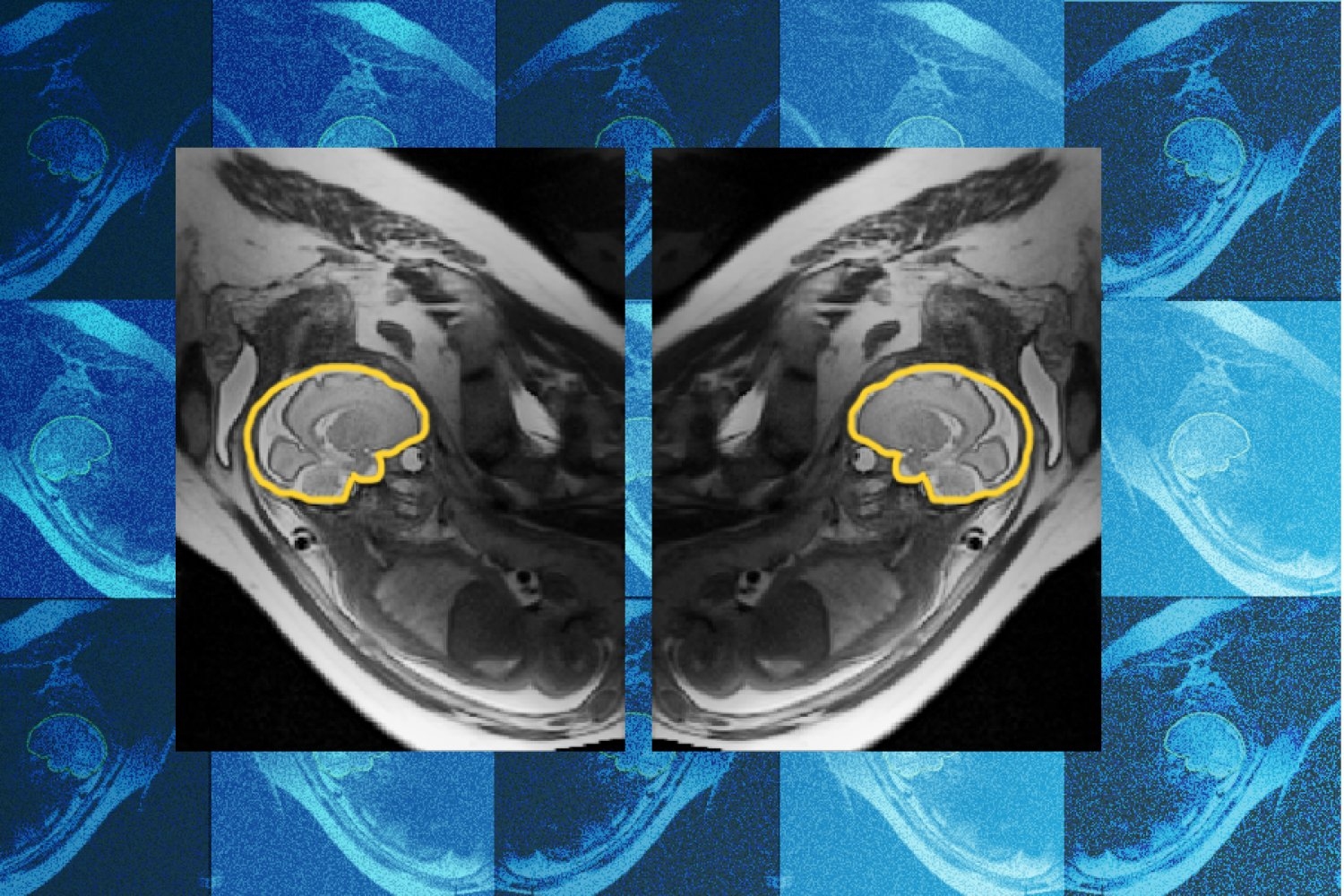
The development of the human fetus is a complex process vulnerable to genetic, environmental, and metabolic disruptions. These disruptions can result in developmental abnormalities, including brain abnormalities. During prenatal ultrasounds, sonographers flag possible brain abnormalities for further magnetic resonance imaging (MRI).
However, imperfect imaging can lead to missed or incorrect diagnoses. To assist in early detection and treatment of these abnormalities, researchers from MIT, Boston Children’s Hospital, and Inria have created and trained a deep learning model named E(3)-Pose.
A paper about the work was presented at the 2026 European Conference on Computer Vision. One challenge with fetal MRI scans is that the baby's movements cause the slices to be irregular, resulting in gaps and unusual orientations. This can lead to missed or incorrect diagnoses by radiologists. E(3)-Pose helps address this issue by focusing on physics, specifically the left-right symmetry of the fetal head. This symmetry is embedded in the model architecture, making it easier to accurately identify the brain's orientation.
Ramya Muthukrishnan, the first author of the paper and an MIT PhD student, began working on the project with no background in physics. She recalls the challenges of implementing the model on the scanner, often resulting in crashes. However, her perseverance led to the development of a sequence that acquires additional MRI scans to improve training and troubleshoot the scanner.
Co-authors Borjan Gagoski and Aryn Lee, along with senior authors Elfar Adalsteinsson, Ellen Grant, and Polina Golland, contributed to the project's success.
Golland notes that Muthukrishnan's work combines geometric concepts like equivariance and symmetry with deep learning to change MRI image acquisition and improve radiologists' ability to detect and treat fetal abnormalities. The applications of E(3)-Pose could extend to other hard-to-image anatomy and even autonomous driving. Muthukrishnan hopes to improve the model to assist with imaging second-trimester or younger fetuses, as early detection of brain abnormalities is crucial for intervention.
Written by urgent.news from MIT News Research's reporting — not their text. Machine-written — may contain errors; check the original before relying on it.
This story
This is one outlet's version. Read the fullest account.