Brain scans reveal opposite reward responses in ADHD and autism
A new meta-analysis reveals that while ADHD and autism both feature atypical brain responses to rewards, the underlying neural activity goes in completely opposite directions, providing biological clues to their distinct behavioral symptoms.
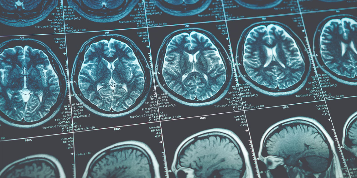
Attention deficit hyperactivity disorder (ADHD) and autism spectrum disorder (ASD) both exhibit atypical reactions to rewards, yet these reactions occur in contrasting ways. A recent study, published in Molecular Psychiatry, reveals that while both conditions involve similar deep brain structures, the patterns of increased or decreased activity differ significantly.
Understanding these differences could shed light on the distinct symptoms associated with each condition. The study, led by researchers from Southwest University in China, analyzed data from 29 existing brain imaging studies involving 468 individuals with ADHD, 424 with ASD, and 1,027 control participants. Functional magnetic resonance imaging (fMRI) was employed to scan participants’ brains while they received either monetary or social rewards.
The researchers utilized specialized software to map brain areas with abnormal activity across the studies. They discovered that both ADHD and ASD involve altered activity in the amygdala and putamen, deep subcortical brain regions involved in processing emotions and motivation. The amygdala, responsible for recognizing motivationally important stimuli, and the putamen, which gauges reward sensitivity, showed opposite patterns of activity.
In ADHD, these areas exhibited abnormally high activity during reward delivery, while in ASD, they showed unusually low activity compared to controls. Additionally, the cortex, the brain's outer layer, displayed distinct changes in both conditions. Individuals with ADHD showed reduced activity in the prefrontal cortex and other areas crucial for impulse control and decision-making, whereas those with ASD displayed heightened activity in medial prefrontal regions associated with assigning subjective value to specific interests.
To gain further insights, the researchers conducted a second analysis using an independent database of healthy brain scans. They examined how the altered amygdala and putamen regions functionally connected with other brain regions. Regions that were hyperactive in ADHD were linked to circuits driving motivation and emotional responses, while those that were hypoactive in ASD were more strongly connected to social and cognitive networks.
Furthermore, the researchers used the Neurosynth database, which contains data from thousands of published studies, to correlate the abnormal brain maps with specific cognitive functions. The hyperactive regions in ADHD matched terms related to intense emotions, while the hypoactive regions aligned with attention and executive control.
For ASD, hyperactive regions were linked to social and value-related terms, whereas hypoactive regions matched novelty and basic affective processing. These findings suggest that reward responses in ADHD are linked to emotional regulation challenges, while ASD responses point to difficulties in integrating social and affective signals.
The study also compared the brain activity maps with known distributions of dopamine and serotonin receptors in the brain. Dopamine, involved in immediate reward-seeking behavior, and serotonin, which manages long-term goals and impulse inhibition, were more densely located in the hyperactive regions of ADHD. In contrast, the hypoactive regions in ASD were rich in these receptors, indicating a general biological desensitization to rewards.
These biological differences provide a framework for understanding the divergent behaviors seen in ADHD and ASD. However, the study acknowledges its limitations and the need for further research.
Written by urgent.news from PsyPost's reporting — not their text. Machine-written — may contain errors; check the original before relying on it.