Standard brain scans might misread oxygen metabolism in large portions of the cortex
A recent brain imaging study reveals that standard fMRI scans can yield backward metabolic readings in nearly half of responding brain regions, challenging conventional interpretations of how neural activity is mapped.
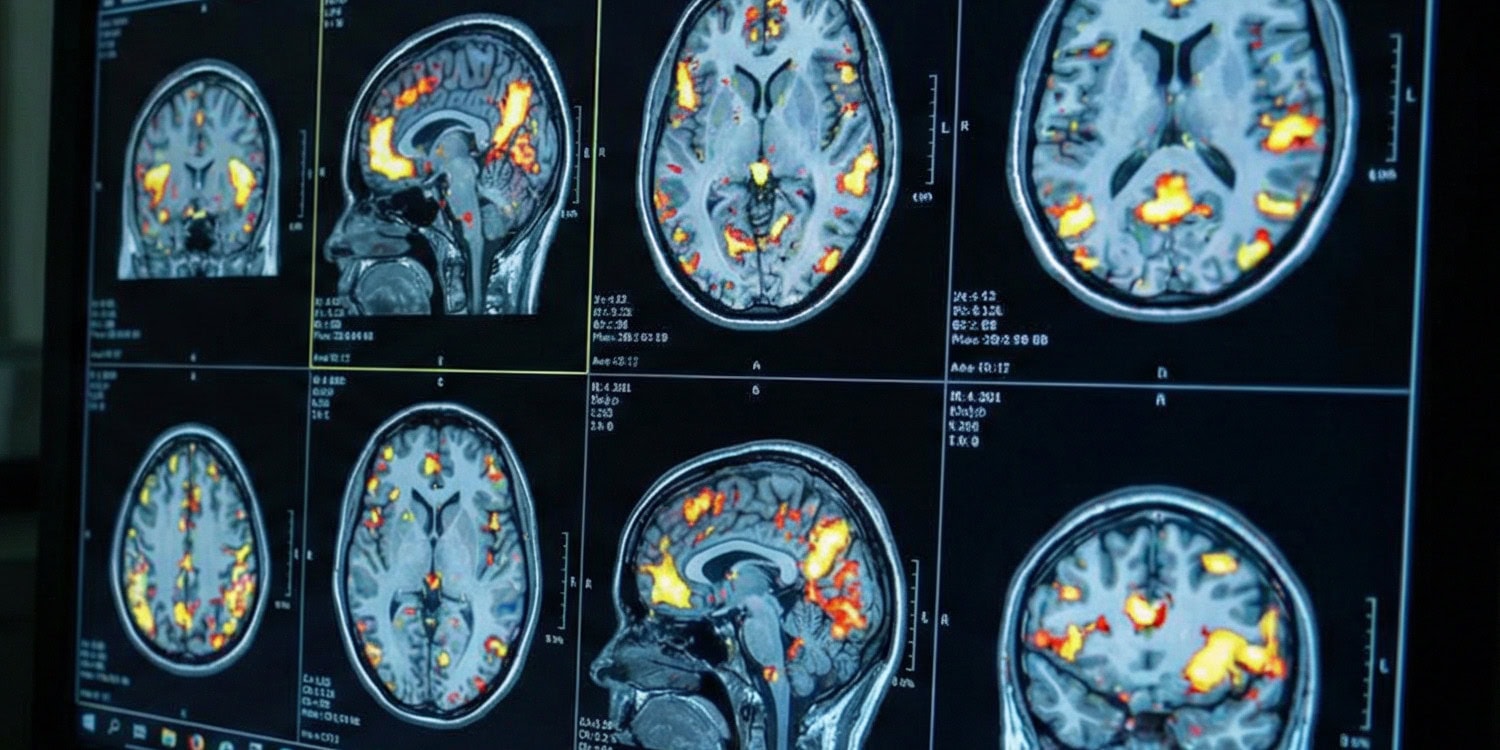
A small study indicates that traditional brain scans, specifically functional magnetic resonance imaging (fMRI), may often produce inaccurate readings of oxygen metabolism in certain brain areas. This finding challenges the conventional interpretation of brain imaging results. Functional magnetic resonance imaging determines brain activity by detecting changes in blood oxygen levels, known as the blood-oxygenation-level-dependent (BOLD) signal.
The prevailing understanding posits that an increase in the BOLD signal signifies heightened local brain activity, while a decrease suggests reduced activity. However, this study reveals that this relationship may not hold uniformly across the entire brain. The research, published in Nature Neuroscience, involved 40 healthy participants who performed various mental tasks inside an MRI scanner.
By comparing the BOLD signal with actual oxygen consumption, the researchers identified anomalies in 40 percent of the brain voxels. In some cases, the BOLD signal indicated heightened activity, yet oxygen consumption decreased. Conversely, in other instances, a negative BOLD signal suggested increased oxygen metabolism. This discrepancy implies that standard brain scans could misinterpret the energy consumption of specific brain regions, particularly those activated by distinct cognitive tasks.
The study underscores the need for more nuanced interpretations of brain imaging data to accurately assess brain function.
Written by urgent.news from PsyPost's reporting — not their text. Machine-written — may contain errors; check the original before relying on it.