High-speed microscopy reveals electrical activity across the brain
The new technique could help scientists learn how the entire brain works to generate decisions and emotions.
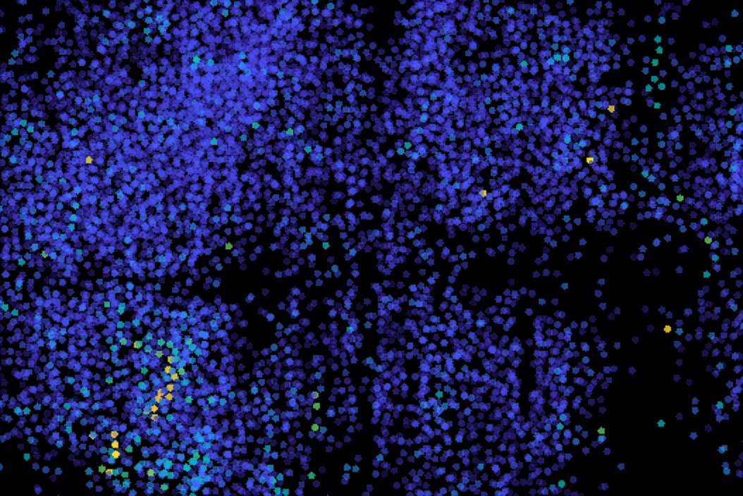
Neurons communicate through electrical impulses, forming complex networks that govern various brain functions. A team of MIT engineers has developed a novel high-speed microscope capable of capturing the electrical activity of neurons distributed across an entire organism, specifically the zebrafish model Danio rerio. This breakthrough, described in a paper published in Nature Methods, could significantly advance our understanding of how neural activity underlies behavior and other brain functions.
Traditionally, calcium imaging has been used to measure neuron activity, as calcium levels increase following an electrical impulse. However, this method is slow, providing only activity data on the order of seconds or minutes. Voltage imaging, on the other hand, offers direct observation of neural activity, enabling high-speed analysis of electrical impulses.
To achieve this, the researchers adapted a commonly used light sheet microscope, a technique that illuminates a thin slice of a sample using a sheet of laser light. By imaging multiple layers in sequence, this method can generate 3D images of large volumes. The team modified this microscope to enhance image acquisition speed and scanning speed, allowing them to scan the entire zebrafish brain at a rate of 200 times per second, or once every five milliseconds.
The researchers engineered zebrafish neurons to express a voltage indicator called Positron2-Kv. Although the indicator did not light up in every neuron, about one quarter of the neurons exhibited acceptable signals, allowing the team to observe patterns of activity across the brain. They imaged the brain while the fish were resting and found that they could detect single voltage spikes from neurons, as well as rapid bursts of spikes.
Additionally, the technique revealed patterns in brain activation following a stimulus, such as ultraviolet light. In response to the stimulus, activity was observed in the optic tectum, a brain region responsible for processing visual input. This activity propagated across a part of the brain called the tectum, demonstrating the interconnected nature of brain regions.
The researchers also noted stimulus-independent activity patterns in sets of neurons throughout the cerebellum and hindbrain.
The advancement of this high-speed microscope opens up new possibilities for neuroscientists to study the brain's complex network of neurons, providing a more comprehensive understanding of how electrical impulses contribute to various brain functions and behaviors. The team plans to increase the percentage of neurons imaged across the brain and improve the microscope's speed and resolution, as well as expand the use of this technique to other experimental models, such as mice.
Written by urgent.news from MIT News Research's reporting — not their text. Machine-written — may contain errors; check the original before relying on it.