Q&A: Lowering radiation dose in chest X-ray imaging yields same accuracy, researcher finds
Digital tomosynthesis (DTS) is an X-ray technique that provides sectional images with better lesion visibility than conventional chest radiography (CXR), while using far less radiation than CT. Masoud Jadidi will defend his thesis, "Evaluation of image quality, radiation dose, and tumor size assessment in chest tomosynthesis," on Aug. 21, 2026. His thesis evaluates digital thoracic tomosynthesis…

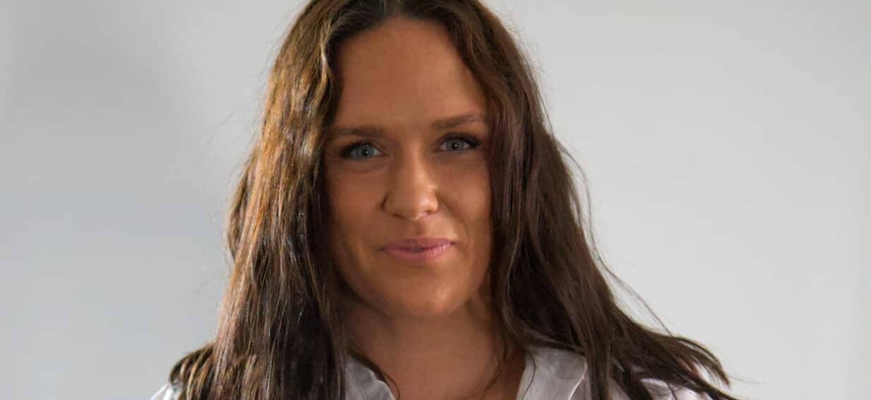
Digital tomosynthesis (DTS) allows for chest X-ray imaging with enhanced lesion visibility, while consuming a fraction of the radiation compared to conventional chest radiography (CXR) and computed tomography (CT). Masoud Jadidi, a doctoral student in the Diagnostic Radiology group, will present his thesis on Aug. 21, 2026, which evaluates DTS as a low-dose method in thoracic radiology.
Jadidi's research examines how image quality is impacted by varying acquisition and reconstruction protocols, as well as potential radiation dose reductions without sacrificing clinical image quality. The findings reveal that optimized DTS protocols, including shorter exposure times and up to 30% dose reduction, maintain high image quality. Additionally, measurements of lung tumors/lesions and consolidations with low-dose DTS exhibit high accuracy and excellent interobserver agreement when compared to CT.
This study supports DTS as a cost-effective, accessible, and low-dose alternative for diagnosing, monitoring, and potentially screening lung cancer. By reducing patients' radiation exposure, improving access to advanced lung imaging, and enabling gentler and more cost-effective follow-up examinations, DTS can contribute to better survival rates in lung diseases, promote equitable healthcare, and optimize limited healthcare resources.
The technique's compatibility with existing X-ray equipment and minimal resource requirements make it particularly beneficial for low- and middle-income countries, where CT capacity and costs pose significant barriers to early diagnosis and follow-up.
Jadidi's future objectives include refining and promoting the use of optimized, low-dose digital chest tomosynthesis in thoracic radiology, both through clinical implementation and methodological development. He aims to integrate DTS into lung tumor/lesion and consolidation follow-ups, as well as future lung cancer screening programs, to ensure more patients receive safe, cost-effective, and accessible diagnostic imaging with reduced radiation doses.
Moreover, Jadidi intends to contribute to international guidelines and education, ensuring the standardized and evidence-based use of this technique, particularly in low- and middle-income countries where the need for resource- and dose-saving methods is most critical.
Written by urgent.news from Medical Xpress's reporting — not their text. Machine-written — may contain errors; check the original before relying on it.